Imaging of Matter
X-rays show self-organization of nanocrystals live
23 May 2019

Photo: DESY, Irina Lokteva
Using X-rays, a research team has watched live how nanocrystals organize themselves into a highly ordered layer. The study helps to better understand the self-organization of nanoparticles, which is important for many technical applications, for example in electronics and optoelectronics, as the scientists write in the journal Small.
Nanocrystals are known to form ordered structures, for example during the controlled evaporation of a solvent. These so-called superstructures can develop completely new material properties through the interaction of the many individual crystals. “However, the result of this self-organisation of the crystals is difficult to predict and even more difficult to control,” explains DESY researcher Irina Lokteva, main author of the publication, who is a member of the cluster of excellence “CUI: Advanced Imaging of Matter”.
In order to better understand self-organization, the scientists investigated lead sulfide nanocrystals as a model system. The individual crystals typically had a diameter of just under four nanometers (millionths of a millimeter). “Lead sulfide is used in solar cells, light-emitting diodes and transistors,” says Lokteva, who works in the DESY research group Coherent X-ray Scattering of Prof. Gerhard Grübel (Universität Hamburg, DESY) and Felix Lehmkühler (DESY). To prevent the nanocrystals from sticking together during production, they were stabilized with oleic acid, which wraps around the particles - like gummy bears are waxed to prevent them from sticking together. Stabilization with oleic acid is an established process in nanotechnology.
For the time-resolved investigation, the nanocrystals were suspended in the organic solvent heptane. 25 microliters (millionths of a liter) of the suspension were dropped into a sample holder 7 millimeters high and just 0.5 millimeters wide, held in a vacuum. As the solvent evaporated slowly during about two hours, the scientists could watch how a 3D highly ordered layer of nanocrystals formed at the walls of the sample holder from the top to the bottom.
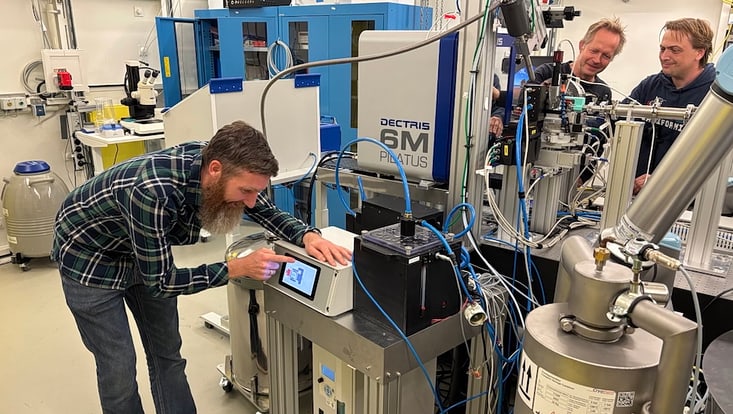
Experimenting at the European Synchrotron Radiation Facility ESRF in Grenoble, the scientists analyzed the exact structure layer every three seconds with X-rays. It turned out that the nanocrystals were initially organized in the hexagonal close-packed (hcp) structure. “We observed this structure for the first time in lead sulfide,” reports Lokteva. However, as soon as the solvent had completely evaporated, the layer changed abruptly into the body-centered cubic (bcc) structure.
“Suddenly, the entire layer jumps into a different structure,” says Lokteva. “Since this is accompanied by a change in volume, cracks appear in the layer.” This effect, which the team observed live for the first time, can either be used specifically or avoided in order to produce layers with desired properties. “With the right framework conditions for self-organization, the final structure can be influenced according to the requirements of the application,” explains Lokteva.
The observation of the lead sulfide model system also yields important insights for other materials. “Numerous applications of nanoparticles require ordered films,” says Lokteva. The research results are a step towards better understanding both the self-organization of nanoparticles and the interaction of the oleic acid ligands with the organic solvent heptane. Text: DESY, ed.
Reference:
Irina Lokteva, Michael Koof, Michael Walther, Gerhard Grübel und Felix Lehmkühler
“Monitoring Nanocrystal Self-Assembly in Real Time Using In Situ Small-Angle X-Ray Scattering”
Small, 2019


