CUI: Advanced Imaging of Matter
Cluster of ExcellenceCUI: Advanced
Imaging of Matter
Imaging of Matter
Photo: UHH/Denstorf
16 July 2025
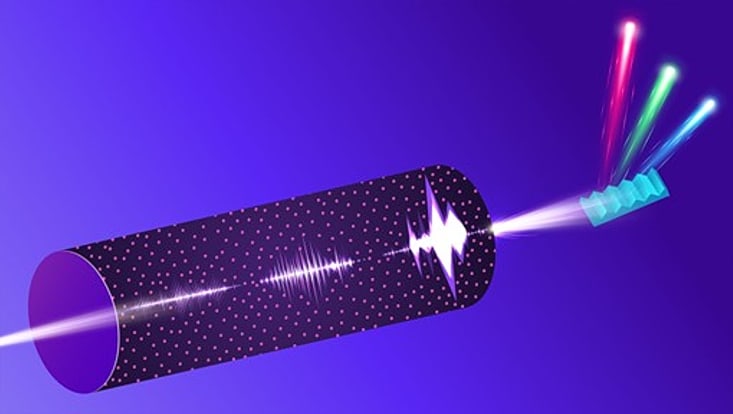
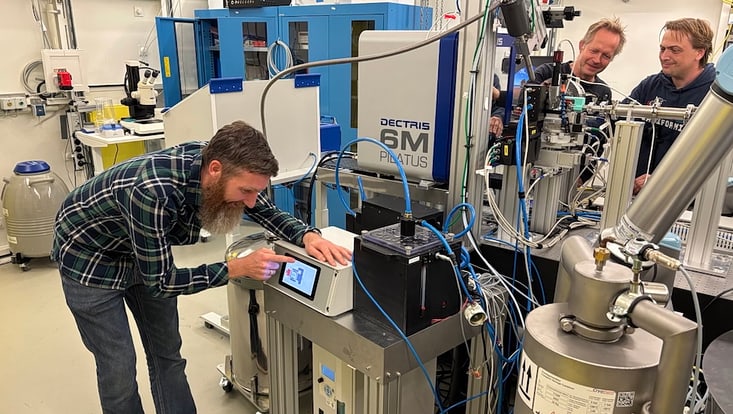
Photo: Stacy Huang/Argonne National Laboratory
A new method turns noise into valuable data to enhance understanding of chemical reactions and material properties with unprecedented detail on atomic level. The results of this research were now published in Nature.
In a leap forward for atomic-scale imaging, researchers have introduced a novel X-ray technique that could transform our understanding of electron motion at the microscopic level. This cutting-edge method, developed by an international team of scientists, uses the unique properties of the X-ray laser of European XFEL at Schenefeld near Hamburg, Germany, — the largest X-ray laser in the world — to capture detailed snapshots of atomic interactions.
The technique, called stochastic Stimulated X-ray Raman Scattering (s-SXRS), turns noise into valuable data, offering snapshots of the electronic structures of atoms. This advancement sets the stage for breakthroughs in chemical analysis and materials science.
Researchers from the U.S. Department of Energy's (DOE) Argonne National Laboratory, the Max Planck Institute for Nuclear Physics, the Small Quantum Systems (SQS) scientific instrument of European XFEL and others developed this innovative approach to X-ray spectroscopy, achieving unprecedented detail and resolution.
“For a long time, chemists have dreamed of seeing how electrons move when they're in excited states, as these movements are what drive chemical reactions,” says Linda Young, an Argonne Distinguished Fellow and professor at the University of Chicago. “Our technique brings us closer to realizing that dream.”
The key innovation is a super-resolution technique that greatly improves the detail in X-ray spectroscopy, a method for studying electron placement around atomic centers. This advancement helps scientists identify closely spaced energy levels in atoms, offering a clearer view of their electronic structures, which determine chemical properties.
“Think of it like upgrading from a standard-definition television to an ultra-high-definition screen,” Young explains. “We're now able to see the fine details of electronic motion that were previously blurred or invisible.”
The practical applications of stochastic Stimulated X-ray Raman Scattering are wide-ranging. For example, it can provide insights into how chemical bonds form or break, offering a deeper understanding of fundamental processes relevant to chemical analysis. This knowledge is essential for developing new materials with specific electronic properties, impacting industries like electronics and nanotechnology.
The researchers directed the X-ray pulses of European XFEL through neon gas and used a spectrometer to collect the resulting radiation. The small, 5-millimeter gas cell was designed by the Max Planck Institute for Nuclear Physics The intense beam created tiny holes in the cell’s entrance and exit windows, allowing the X-rays to pass through to a grating spectrometer — a device that separates light into its different wavelengths — provided by collaborators from Uppsala University in Sweden. The European XFEL experts have taken on a vital role in coordinating the installation and performing thorough pre-experimental testing. “This ensured optimal focusing conditions, which were crucial for efficiently acquiring a large amount of data during the experiment” explains Michael Meyer, group head of the Small Quantum Systems (SQS) instrument at European XFEL and a researcher in the Cluster of Excellence 'CUI: Advanced Imaging of Matter'.
As the X-rays pass through the gas, they amplify the Raman signals — a type of X-ray fingerprint that provides information about the excited electronic states of atoms or molecules — by nearly a billion-fold. This amplified signal provides detailed information about the electronic structure of the gas on a femtosecond timescale, or one quadrillionth of a second. By analyzing the relationship between the incoming pulses and the resulting Raman signals, scientists can create a detailed energy spectrum from many individual snapshots, rather than scanning slowly across different energy levels.
“The large number of pulses in each X-ray flash not only boosts the measurement signal but also holds the key to the highest spectral resolution by averaging over many photon impacts on the detector at once,” says Thomas Pfeifer from the Max Planck Institute for Nuclear Physics and the Cluster of Excellence "STRUCTURES" at the University of Heidelberg
“This approach, pinpointing the center position of broad but distinct spectral spikes much more precisely than the width of the spikes, is similar to the super-resolution microscopy technique that won the 2014 Nobel Prize in chemistry”, Pfeifer adds.
The stochastic Stimulated X-ray Raman Scattering uses a statistical method, called covariance analysis, to link the incoming X-ray pulses with the emitted Raman signals. This transforms what was once considered “noise” into a valuable resource, allowing extraction of detailed information from complex data. This approach not only enhances the resolution, but also speeds up data collection, providing rapid and detailed snapshots of atomic interactions.
Another crucial part of this research involved the Argonne Leadership Computing Facility (ALCF), a DOE Office of Science user facility, which provided the necessary computational power to simulate the complex interactions between X-ray pulses and matter. These simulations were vital for interpreting the experimental results and refining the technique.
The detailed simulations closely matched the experimental data, providing valuable insights into how X-ray pulses behave as they travel through gases. The computations were instrumental in confirming the researchers’ understanding of how the X-ray pulses move and interact, paving the way for future investigations.
With continued advancements, s-SXRS could become a standard tool in laboratories worldwide, driving innovation across many fields.
Michael Meyer is convinced: “This study is an excellent example of the capability of the European XFEL, especially of its high intensities and the recently demonstrated generation of extremely short X-ray pulses with durations of less than one femtosecond. These advancements will certainly trigger further investigations to unravel the dynamics of complex chemical reactions.”
“We’re just beginning to scratch the surface of what we can achieve with this level of detail,” Young says. “It's an exciting time for science and technology.” Text: XFEL, ed.
Other contributors to this work include Christian Ott, Alexander Magunia and Marc Rebholz from the Max Planck Institute for Nuclear Physics; Marcus Agåker from Uppsala University and Lund University; Phay Ho and Gilles Doumy from Argonne National Laboratory; Marc Simon from Sorbonne University; Tommaso Mazza, Alberto De Fanis, Thomas M. Baumann, Jacobo Montano, Nils Rennhack, Sergey Usenko, Yevheniy Ovcharenko from European XFEL; Kalyani Chordiya from the University of Hamburg and Louisiana State University; Lan Cheng from Johns Hopkins University; Jan-Erik Rubensson from Uppsala University; and Mette B. Gaarde from Louisiana State University.
This study was funded by the DOE Office of Science, Basic Energy Science, Chemical Sciences, Geosciences and Biosciences Division and the Hamburg Cluster of Excellence ‘CUI: Advanced Imaging of Matter’ and the Heidelberg Cluster of Excellence "STRUCTURES" of the DFG, Germany, the European Research Council (ERC) and the Max Planck Society.
K. Li, C. Ott, M. Agåker et al.
Nature 643, 662–668 (2025)