CUI: Advanced Imaging of Matter
Cluster of ExcellenceCUI: Advanced
Imaging of Matter
Imaging of Matter
Photo: UHH/Denstorf
20 August 2026
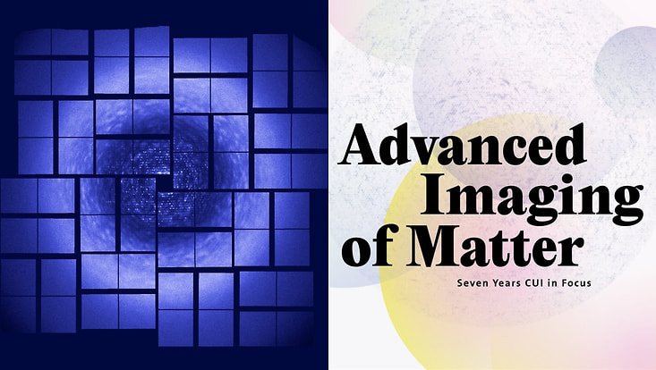
Photo: DESY, O. Yefanov, UHH/DESY, H. Chapman, ASU, P. Fromme (left); UHH/CUI (right)
When a bunch of electrons travels at near the speed of light through a 100-meter long periodic array of magnets after being accelerated to giga-electron-volts in a linear particle accelerator and compressed into a pulse only femtoseconds in duration, it radiates the brightest flashes of X-rays ever made. In the magazine “Advanced Imaging of Matter – Seven Years CUI in Focus,” Prof. Henry Chapman provides an overview of the progress that has been made in research using X-ray lasers.
The process described above is known as self-amplification of spontaneous emission. It happens thousands of times per second in the European XFEL in Hamburg as well as at other X-ray free-electron lasers throughout the world, and relies on exquisite tuning and precision along the entire length of the kilometers-long facility. The electrons are driven into correlated oscillations by a continuous inter-action with the intense light they generate – a positive feedback that creates spatially coherent X-ray light. With peak powers billions of times higher than at synchrotron radiation facilities, these sources have opened up a vibrant field of ultrafast X-ray science. Scientists at CUI have been at the forefront of developing and applying methods that utilize this radiation to explore the structure and dynamics of materials at the length-and timescales of atoms.
One of the first things an experiment at an XFEL has to contend with is that the high intensity of the pulses vaporizes any material that is placed in the path of the focused beam. However, such destruction evolves only after the femtosecond pulse has passed through the sample and is not usually apparent in the diffraction pattern or spectrum that is generated. The X-ray pulse out-runs the damage. This method of “diffraction before destruction” has opened up the possibility of determining protein structures from samples that are too radiation-sensitive to be measured at conventional X-ray sources, and has eliminated the need to cryogenically cool macromolecular crystals.
However, as the sample is destroyed in a single shot, this has required a new paradigm for measuring three-dimensional structures. In this approach, called serial crystallography, a stream of small crystals is sped across the beam in a liquid jet, an aerosol, or by some other means. At the European XFEL, more than 3,000 individual fresh new crystals can be measured each second, limited by the speed of the detector. The rapid stream of data can be merged after first figuring out the orientation of each crystal to build up a three-dimensional map in the frame of reference of the crystal lattice.
Because the exposure time is so short, the method enables the measurement of 3D structures as a function of time. Photoactive proteins can be triggered by a flash of light from an optical laser that is precisely synchronized to arrive just before the X-ray pulse. For example, the relative delay was scanned to build up frames in a “molecular movie”, to follow the trans-to-cis isomerization of the chromophore in the photoactive yellow protein (PYP), which occurs less than 500 femtoseconds after photo-absorption. The temporal resolution in such experiments is limited to about 100 femtoseconds by the jitter in the synchronization between the excitation or “pump” pulse and the X-ray “probe”, but a powerful machine-learning approach has been able to sort the millions of patterns collected into a time series on a much finer scale approaching 10 femtoseconds. In this way, Prof. Robin Santra and his colleagues observed the structure transition through a conical intersection of the potential-energy surfaces of the excited and the ground state, supported by a Fourier analysis which reveals the same frequencies as observed by Raman spectroscopy. This demonstrates the power of these big data experiments to extract rare and transient events in a process.
Photoactive proteins are certainly relevant to the processes of photosynthesis and vision, and recent work in CUI has examined light-driven proteins that repair DNA. But how can we study the dynamics of the vast majority of macromolecules that do not respond to light? One way is to engineer light triggers, such as compounds that sever upon light absorption to free a reactant that quickly diffuses to an active site of a protein. Prof. Arwen Pearson has been devel-oping these “photocages” to initiate reactions for experiments at XFELs and synchrotron sources. Over longer timescales of milliseconds, mixing of a reactant solution with the slurry of protein crystals allows intermediate states to be captured in reactions of drug-resistant strains of bacteria that cause tuberculosis. These show the protein adapting to accept the compound, giving hints on how to better adapt the drug. Serial crystallography has opened up a new field of macromolecular structural dynamics, where time-dependent structural landscapes provide a new understanding of how proteins function.
The high pulse rates of the European XFEL are enabling the serial measurement approach to be extended to nanoparticles and single macromolecules. Even with the extreme intensities of the XFEL, objects as small as a single protein give very weak measurable diffraction signals, which can be lost in background noise. Many millions of patterns are required to build up a signal above this noise, but each pattern has to be first oriented into a common frame of reference. Without a crystal lattice to give a recognizable pattern, this step has proven to be challenging. A proof-of-principle experiment was carried out on gold nanoparticles, where it was possible to acquire 10 million individual patterns. Machine learning provided the means to combine the weak patterns into a latent space that could track the 3D structures under transition.
This approach is now being applied to time-resolved studies of the coupling of light to nanostructures, such as polariton dynamics. Prof. Jochen Küpper and his colleagues in CUI have developed instruments to produce fine beams of single particles from aerosols, to deliver them into the X-ray focus. His group is also perfecting an alternative approach that extracts structural information from an ensemble of molecules, by aligning all the molecules in space and even selecting quantum states, so that diffraction patterns can be accumulated over many pulses. Yet another method pioneered in Hamburg is based upon Coulomb ex-plosion imaging (see p. 46 in the magazine), where the atomic fragments of molecules are detected in an ion momentum spectrometer. By using intense XFEL pulses to simultaneously ionize multiple atoms in a single molecule, a very clear localization of ions can be obtained from the exploding molecule, which can be used to recover structures and to follow charge rearrangements and other ultrafast molecular dynamics, molecule by molecule.
In addition to zooming into single molecules, XFELs are used in CUI to explore the many-body physics of ensembles to study the structural dynamics of liquids and glasses. By measuring the correlations in diffraction patterns, order parameters, diffusion dynamics, and interaction strengths can be tracked in microscopic volumes and on short timescales. One of the most intriguing liquids is water, whose anomalous properties can now be investigated by correlation scattering measurements of supercooled water jets.
The extreme intensities from XFELs have been used to extend non-linear and quantum optics into the X-ray regime. The first inner-shell laser was demonstrated by Prof. Nina Rohringer, who created a population inversion by rapidly ionizing neon atoms, to produce a spectrally pure and fully coherent X-ray pulse by stimulated emission. Stimulated Raman scattering allows the study of coherent electronic wave packets in catalysts and enzymes, as needed to learn how to control energy-transfer processes in such systems. By coherently mixing optical pulses, CUI’s Young Investigator, Dr. Christina Bömer, brings the capability to probe valence dynamics, responsible for many optical properties of materials, to the atomic scale – a unique capability that will provide insights into the creation of new light-driven functionalities in materials. Femtosecond pulses also enable a new kind of photon-photon correlation imaging demonstrated at CUI, to create chemical-specific atomically resolved images of molecules by interference of fluorescence emission.
X-ray FELs are continuously developed and improved to produce even more intense and shorter pulses, down to the attosecond regime. These will open up further opportunities, ex-tending CUI’s capabilities in femtochemistry to fully explore electron correlations directly in time as they interact with the evolving nuclear arrangements. Indeed, a strength of CUI has been in combining concepts from conventional X-ray analysis with ideas of coherence and control from laser physics, condensed-matter physics, and femtochemistry to exploit the extraordinary properties of XFELs. Text: Henry Chapman (UHH/DESY)